Zackeryeh Salloum – MRes Student, University of Portsmouth

We have awarded a grant to Zackeryeh Salloum, an MRes stduent working in the School of Environment, Geography and Geosciences in Portsmouth.  Zack previously studied Bsc Marine Environmental Science at the University of Portsmouth and completed his final year project on the changing microplastic distribution within Swansea Bay in two varying seasons. This included the use of multiple lab methods in microplastic analysis and identification, from which the further research is being developed. This research will make up the main body of an MRes project, the aim is to publish the final report and develop a new method of microplastics analysis.

About the Research

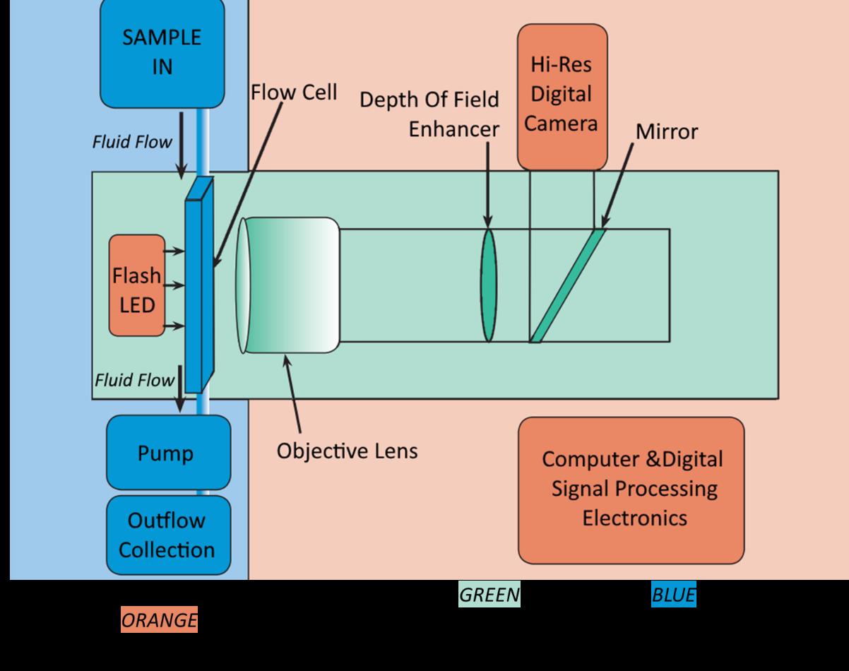
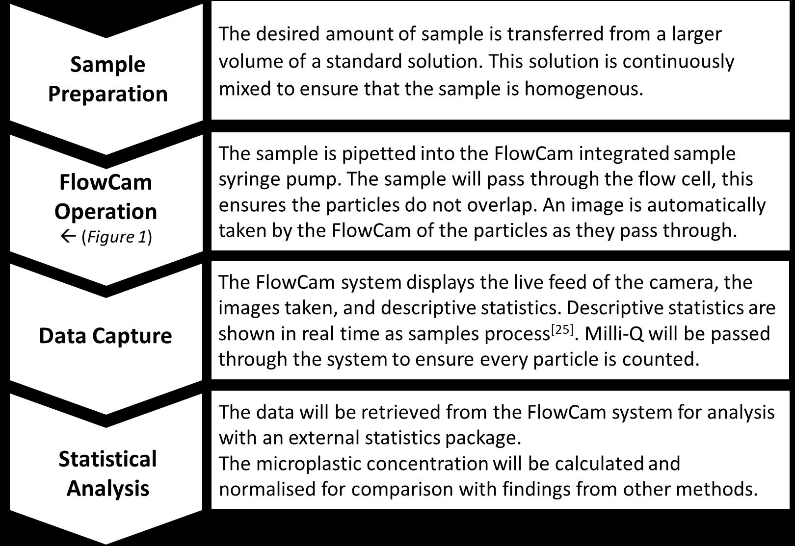
Zack’s research aims to produce a streamlined and accurate method of enumerating microplastic particles using FlowCam technology. This apperatus has been previously used to count cells of phytoplankton as well as provide a microscope image of every individual cell.  By using an adapted method of processing the equipment can be used to image and accurately count microplastics. To test whether the method is reliable and useful it will be compared to multiple current methods such as FTIR analysis and microscope counts.

This study aims to compare multiple methods to a new microplastic numeration method using a FlowCam imaging flow cytometer, which would be beneficial for further studies of microplastics. FlowCam analysis is far quicker than most other methods as high throughput samples can be run rather than taking smaller sub-counts or sub-samples.  The application of FlowCam can provide a cheaper alternative than FTIR as a standalone method, this would be highly useful for industries that are working to lower their emissions of microplastics e.g. wastewater treatment plants. The ability to process samples accurately without needed to run entire sample volumes through FTIR processes make large samples easier to manage, this would allow more vigorous testing of facilities in a similar timeframe. These tests would be highly important to understanding where the sources and sinks for hazardous microplastics are within both industrial systems and environments.

Microplastic Identification and Analysis

Microplastics are “Synthetic solid particles or polymeric matrix, with regular or irregular shape and with size ranging from 1μm to 5mm, of either primary or secondary manufacturing origin, which are insoluble in water”[1]. Microplastics also can transport harmful pollutants such as Polycyclic aromatic hydrocarbons (PAHs) and heavy metals, this increases bioavailability and leads to bioaccumulation within food chains[2 – 6]. Understanding how microplastics effect organisms and ecosystems is crucial for the preservation of marine environments[7,8].

Sample Production and  Preparation

Sample preparations will be taken for all methods, a stock solution is made using premade microplastics and filtered Milli-Q water.  Multiple concentrations will be tested and compared to ensure microplastics can be easily identified across all analytical methods. A specific concentration will be sampled across all methods allowing comparisons of measured microplastic concentrations.

Current methods of Analysis

Light Microscope counts and Nile red stain counts: Samples will be counted using light microscope using a Sedgewick Rafter Chamber. Samples stained with Nile Red will be transferred onto a Millipore filter (2 µm) and examined under fluorescence microscope [9]

Flow cytometry: A flow cytometer will be used to identify microplastics using a method based on multiple studies [10–13]

FTIR: Samples will be run through a Fourier Transform InfraRed spectrometer (FTIR), this is one of the most used methods of microplastic identification due to the high level of accuracy [9,13–16].

Micro-Ramen: Samples will be analysed by a Micro-Raman spectrometer, another highly accurate method of determining microplastics specifically in the smaller ranges (<50 µm) [9,16–18].

FlowCam: Current Applications and Method

The FlowCam VS imaging flow cytometer is used in the identifying and numerating many taxa of phytoplankton and zooplankton [ 19-22], VisualSpreadsheet is then used to automate previously time-consuming microscope counts[23-25]. FlowCam has the potential to process high volumes of sample all in a quick timeframe, which other methods would not be able to do.

FlowCam has frequently been used for the imaging and study of oyster larvae, studies used the FlowCam fluorescence spectroscopy to further investigate samples[26–28]. As microplastic particles are of similar size as plankton, zooplankton, and oyster larvae; a similar method may be taken to investigate microplastic samples.

There are very few examples of the use of FlowCam for microplastic investigation literature, consisting of 2 conference abstracts and one scientific paper. Of the research found none put forth a methodology of study using FlowCam.

References

[1]Frias, J. et al. Mar. Pollut. Bull. 138, (2019).
[2]Avio, C. G. et al. Environ. Pollut. (2015) doi:10.1016/j.envpol.2014.12.021.
[3]Sharma, S. et al. Environ. Sci. Pollut. Res. 24, 21530–21547 (2017).
[4]Botterell, Z. L. R. et al. Environmental Pollution (2019) doi:10.1016/j.envpol.2018.10.065.
[5]Koelmans, A. A. et al. Water Res. 155, 410–422 (2019).
[6] Peng, L. et al. Sci. Total Environ. (2020) doi:10.1016/j.scitotenv.2019.134254.
[7]Cole, M. et al. Marine Pollution Bulletin (2011) doi:10.1016/j.marpolbul.2011.09.025.
[8]Wright, S. L. et al. Current Biology (2013) doi:10.1016/j.cub.2013.10.068.
[9]Renner, G. et al. Curr. Opin. Environ. Sci. Heal. 1, 55–61 (2018).
[10]Andrady, A. L. NOAA Tech. Memo. 54 (2010).
[11]Sgier, L. et al. Nat. Commun. 7, (2016).
[12]Mai, L. et al. Environ. Sci. Pollut. Res. 25, 11319–11332 (2018).
[13] Prata, J. C. et al. TrAC – Trends in Analytical Chemistry (2019) doi:10.1016/j.trac.2018.10.029.
[14]Qiu, Q. et al. Estuarine, Coastal and Shelf Science (2016) doi:10.1016/j.ecss.2016.04.012.
[15]Shim, W. et al. Anal. Methods 9, 1384–1391 (2017).
[16]Cabernard, L. et al. Environ. Sci. Technol. 52, 13279–13288 (2018).
[17]Hanvey, J. S. et al. Anal. Methods 9, 1369–1383 (2017).
[18]Collard, F. et al. Arch. Environ. Contam. Toxicol. 69, 331–339 (2015).
[19]Buskey, E. J. et al. Harmful Algae (2006) doi:10.1016/j.hal.2006.02.003.
[20]Poulton, N. in 237–247 (2016). doi:10.1007/978-1-4939-3302-0_17.
[21]Poulton, N. et al. in Microscopic and molecular methods for quantitative phytoplankton analysis 47–54 (2010). doi:10.1016/j.resp.2011.02.009.
[22]Álvarez, E. et al. J. Plankton Res. 36, 170–184 (2014).
[23]Steele, R. (Carleton University, 2019). doi:10.22215/etd/2019-13774.
[24]Camoying, M. G. et al. Limnol. Oceanogr. Methods 14, 305–314 (2016).
[25]Fluid Imaging Technologies. Fluid Imaging Technologies Inc https://www.meritics.com/pdf/FlowCAM_VS Industrial_Brochure_Meritics.pdf (2011).
[26]Mroch, R. M. et al. J. Shellfish Res. 31, 1091–1101 (2012).
[27]Adams, C. et al. Aquac. Environ. Interact. 11, 521–536 (2019).
[28]Gancel, H. N. et al. Estuaries and Coasts 42, 1558–1569 (2019).

Leave a Reply